SHORT CASE 2
This is an online E log book to discuss our patient's de-identified health data shared after taking his/her/guardian's signed informed consent. Here we discuss our individual patient's problems through series of inputs from available global online community of experts with an aim to solve those patient's clinical problems with collective current best evidence based inputs. This E log book also reflects my patient-centered online learning portfolio and your valuable inputs on the comment box .
65 year old male patient r/o Nakrekal presented to casualty with complaints of
C/O Shortness of breath grade 3 since 4 months .
C/O B/L pedal edema since 4 months .
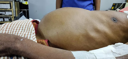
C/O abdominal distension since 5 days
C/O Oliguria since 2 days
HISTORY OF PRESENTING ILLNESS :
Patient is toddy climber by occupation , was apparently asymptomatic 4 months ago .
-Intially he noticed b/l swelling of lower limbs , gradual onset and progressive . Pitting type and extending upto knees .
Associated with shortness of breath grade 2 , progressed to grade 3 over 4 months .
H/O orthopnea and PND present .
No h/o chest pain , palpitations
In view of sob , patient visited local hospital and was told ,he had a stone in one of his kidneys and both his kidneys failed .
He was advised maintainace hemodialysis,but patient denied and was discharged on medications .
His pedal edema subsided after using medications .
He continued taking medications , but noticed loss of appetite, weight , fatigue and generalized weakness .
His urine output was adequate previously . H/o hematuria present occasionally
No h/o pus in urine , burning micturition , frothy urine .
As he had generalized fatigue ,loss of appetite and ,sob ,elevated urea and s. creatinine he visited our hospital and was initiated on hemodialysis by placing central venous catheter in right internal jugular vein .
Patient had 4 sessions of hemodialysis .
He went to Hyderabad and got A-V fistula on his left hand .
C/o Low back ache and body pains .
C/O abdominal distension since 5 days , sudden onset and progressed gradually . Associated with increased sob on lying down and abdominal tightness.
Pedal edema is mild extending upto ankle joint.
No h/o yellowish discoloration of eyes . No h/o binge alcohol intake .
Past history - K/c/o HTN since 10 years and is not on regular medication .
NOT a k/c/o DM, TB , ASTHMA,CAD , EPILEPSY,CVA .
No surgical history and past Medical history
No h/ o NSAID abuse .
Personal history - Regular bowel and bladder movements
Adequate sleep
Loss of appetite present
Mixed diet
Social & Educational History :
Married for 27 years with 2 children. Not educated
Family history - Not significant
Addictions - Toddy drinker occasionally -3 times /week . 90 ml
Non -Smoker
PROVISIONAL DIAGNOSIS :
65 year old male with acute history of oliguria and abdominal distension ,on a background of Sob and pedal edema and HTN
?Acute decompensated Heart failure in view of anasarca and orthopnea and PND .
? Renal failure in view of decreased urine output and Anasarca and h/o renal calculi and HTN
General examination :
Pt C/C/C
Pallor - present
No icterus , clubbing, cyanosis,koilonychia , lymphadenopathy
B/L pedal edema - pitting type present. extending upto ankle .
Jvp - couldn't be assessed due to central line .
Skin - Dry ,scaly , itching present .
Eyes - Grade 2 HTN retinopathy changes noted on fundoscopy .
Vitals :
Bp - 140/90 mmhg - Right arm supine posture
Pulse - 130 bpm ,regular ,normal volume, condition of vessel wall - normal, no radio-radial or radio-femoral delay.
Resp rate - 26/ min
Spo2 - 97% on RA
Grbs - 110 mg/dl
Temp -99 F
SYSTEMIC EXAMINATION :
GIT EXAMINATION :
INSPECTION :
Shape of abdomen - Distended-uniform
Flanks – Full
Umbilicus – Everted
Skin – Stretched, shiny
No scars, sinuses, striae, nodules , discoloration.
Dilated veins – on front present
Movements of the abdominal wall - All quadrants equally moving with respiration .
Abdomino - Thoracic type of breathing
NO visible intestinal peristalsis
Hernial Orifices normal
Cough impulse - Negative
External genitalia - Scrotal swelling present
PALPATION :
Measurements - Abdominal Girth - 108 cm
Flanks - full
Superficial Palpation – Tenderness present in epigastrium
No local rise of temperature
Direction of Blood Flow in Veins - away from umbilicus
Deep Palpation :
Liver Span - Couldn't be palpalted due to gross distension .
Spleen - Couldn't be palpalted due to gross distension
Kidney - Couldn't be palpalted due to gross distension
Any other Palpable swelling - No
Hernial Orifices - normal
Murphy’s Punch/Renal angle tenderness - no tenderness
External Genitalia - scrotal edema present . non tender and trans -luminant
PERCUSSION:
Fluid Thrill - Present
Shifting dullness - Present
AUSCULTATION:
Bowel sounds – Present
Aortic, Renal Bruit - Absent
CARDIOVASCULAR EXAMINATION :
INSPECTION:
Chest wall shape - Ellipsoid and b/l symmetrical
No Precordial bulge, Pectus carinatum/excavatum
No Kyphoscoliosis
No Dilated veins, scars, sinuses
Apical impulse - Visible in left 5 ICS 1 cm lateral to MCL .
Pulsations – epigastric, parasternal - absent
PALPATION:
Apical impulse – Tapping type , felt in left 5 ICS 1 cm lateral to MCL .
Pulsations – No Epigastric pulsations
Parasternal Heave – Present - Grade 2
No Thrills and palpable heart sounds .
Auscultation :
S1 S2 heard in Aortic , pulmonary,tricuspid and mitral areas .
No added sounds
No murmurs
Respiratory system -B/L NVBS
B/L fine crepitations present IAA ,ISA .
CNS - NO abnormality detected .
INVESTIGATIONS :
BGT:- A positive
Serum iron :- 83
CBP :
HB - 7 g/dl
TLC - 12,400 cells /mm3
Platelets -1.3 lakhs .
RFT:-
Urea :- 92 mg/dl
Creatinine :- 5.7 mg/dl
Uric acid :- 6.6 mg/dl
Calcium :- 9.6 mg/dl
Phosphorus :- 3.4 mg/dl
Sodium :- 132 mEq /L
Potassium :- 3.7 mEq/L
Chloride :- 98 mEq/L
RBS:- 79mg/dl
LFT:-
Total bilirubin :- 2.12 mg/dl
Direct bilirubin:- 0.64 mg/dl
AST :- 17 IU/L
ALT :- 10 IU/L
Alkaline phosphatase:- 203 IU/L
Total proteins :-6.1 gm/dl
Albumin:- 2.2 gm/dl
A/G ratio:- 0.56
USG SCANNING OF WHOLE ABDOMEN:-
Impression:-
1) Bilateral GRADE 3 RENAL PARENCHYMAL CHANGES
2) MULTIPLE LARGE LEFT RENAL CALCULI .
3) Moderate to Gross ascitis
COMPLETE URINE EXAMINATION:-
Colour:- pale yellow
Appearance :- slightly hazy
Specific gravity:- 1.010
PH:- Acidic (6.0)
Proteins:- +++
Glucose :- Nil
Urobilinogen:- Negative
Bilirubin:- Negative
Ketones:- Negative
Nitrates :- Negative
Pus cells:- 5-6 /HPF
Rbc:- 5-7 /HPF
Epithelial cells:- 1-2/HPF
Casts:- Granular casts present
Crystals :- Nil
2D echo :-
Impression -
EF:- 48%
Dilated LA
Conc LVH
Moderate MR/ Mild AR / Mild TR
No PAH/ No PE/ No CLOT
No RWMA
Sclerotic AV .
CT KUB : showing left kidney 7-8 cm Staghorn calculus causing mild hydronephrosis and thining of parenchyma
Ascitic fluid analysis showed - HIGH SAAG
Examination videos links :
https://youtu.be/FDq1rKdkIzQ
https://youtu.be/SXvrJfDQzig
https://youtu.be/p_W68yJ8Yu8
Final diagnosis -
Ascitis secondary to portal hypertension - ? Post hepatic -
Secondary to Congestive cardiac failure (HFPEF)
Post renal AKI on CKD - Left staghorn calculus
CKD - Stage 5 - Native kidney disease - ? Htn nephropathy
Cardio- renal syndrome type 4


Comments
Post a Comment